
Label parts of microscope worksheet provides an in-depth exploration of the fundamental components of a microscope, empowering learners with a comprehensive understanding of its structure and functionality. This guide delves into the intricacies of each component, unveiling their roles in magnifying and illuminating specimens, making it an indispensable resource for students and microscopy enthusiasts alike.
As we embark on this journey of discovery, we will dissect the microscope’s anatomy, examining the eyepiece, objective lenses, stage, base, condenser, diaphragm, and illumination system. Through detailed descriptions, illustrative diagrams, and engaging discussions, we will unravel the mysteries of microscopy, empowering you to master the art of specimen observation.
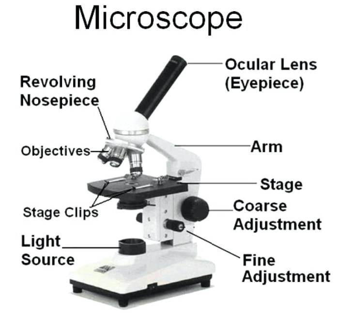
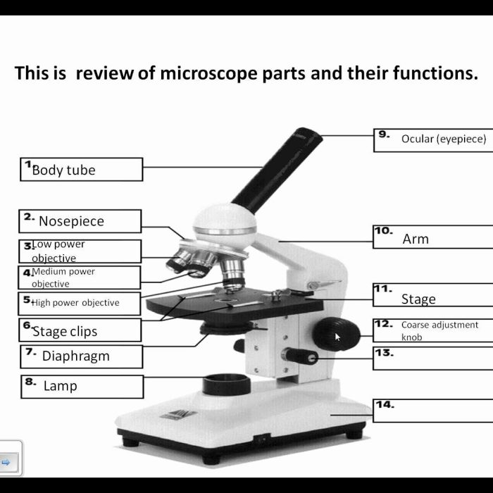
Overview of Microscope Parts
A microscope is a scientific instrument that produces magnified images of small objects. It consists of several essential components, each playing a specific role in magnifying and focusing the image.
The primary components of a microscope include:
- Eyepiece: The eyepiece, also known as the ocular lens, is located at the top of the microscope and is where the observer looks through to view the specimen.
- Objective lenses: The objective lenses are located at the bottom of the microscope and are responsible for magnifying the specimen. Different objective lenses provide different levels of magnification.
- Stage: The stage is the platform where the specimen is placed for viewing. It can be moved up and down to focus the image.
- Base: The base is the supporting structure of the microscope and provides stability.
The following table provides a summary of the microscope parts and their functions:
| Part | Function |
|---|---|
| Eyepiece | Provides magnification for viewing the specimen |
| Objective lenses | Magnify the specimen |
| Stage | Holds the specimen for viewing |
| Base | Provides support and stability for the microscope |
Eyepiece
The eyepiece, also known as the ocular lens, is the lens closest to the observer’s eye. It plays a crucial role in magnifying the image formed by the objective lens, providing the final magnified view of the specimen.
Eyepieces come in different types, each with its own characteristics. Two common types include:
Huygens Eyepiece
- Consists of two plano-convex lenses separated by a short distance.
- Provides a wide field of view and good image quality.
- Used in dissecting microscopes and other applications where a wide field of view is desirable.
Ramsden Eyepiece
- Comprises two plano-convex lenses separated by a larger distance.
- Offers a narrower field of view but higher image resolution.
- Suitable for high-magnification applications, such as compound microscopes.
The illustration below shows the key parts of an eyepiece:
[Insert illustration of an eyepiece with labels: barrel, lens elements, field diaphragm]
Objective Lenses
Objective lenses are crucial components of a microscope, responsible for magnifying the specimen. They are mounted on the revolving nosepiece and can be interchanged to achieve different levels of magnification.
Types of Objective Lenses
There are three main types of objective lenses, each with distinct characteristics and uses:
- Low-power objective lenses:Typically have magnifications ranging from 4x to 10x. They provide a wide field of view, making them ideal for initial examination of specimens and scanning large areas.
- High-power objective lenses:Offer higher magnifications, ranging from 40x to 100x. They provide a narrower field of view but allow for detailed observation of smaller structures within the specimen.
- Oil immersion objective lenses:Have the highest magnification, typically 100x. They are designed to be used with a drop of immersion oil between the lens and the specimen. This reduces light refraction and improves image resolution.
Comparison of Objective Lenses
The following table compares the magnification and resolving power of different objective lenses:
| Objective Lens | Magnification | Resolving Power |
|---|---|---|
| Low-power | 4x
|
0.2
|
| High-power | 40x
|
0.05
|
| Oil immersion | 100x | 0.002
|
The resolving power of a lens refers to its ability to distinguish between two closely spaced points in the specimen. Higher magnification does not necessarily lead to higher resolving power, as other factors such as the wavelength of light and the numerical aperture of the lens also influence resolution.
Stage

The stage is a platform on which the specimen is placed for observation under the microscope. It is typically made of metal or plastic and has a hole in the center to allow light to pass through the specimen. The stage can be moved up and down, left and right, and rotated to position the specimen correctly for viewing.
Types of Stages, Label parts of microscope worksheet
There are two main types of stages: fixed and mechanical.
- Fixed stagesare permanently attached to the microscope and cannot be moved. They are typically used for simple observations of stationary specimens.
- Mechanical stagesare mounted on a rack and pinion mechanism that allows them to be moved precisely in the x and y directions. This type of stage is used for more complex observations, such as when the specimen needs to be moved around to find a specific feature.
Diagram of a Stage
The following diagram shows a typical stage with its key parts labeled:
[Image of a stage with labels]
- Stage clips: These clips hold the specimen in place on the stage.
- Stage control knobs: These knobs are used to move the stage up and down, left and right, and rotate it.
- Aperture diaphragm: This diaphragm controls the amount of light that passes through the specimen. It can be opened or closed to adjust the brightness of the image.
Base
The base is the foundation of the microscope, providing stability and support for the entire structure. It typically consists of a heavy, weighted platform that ensures the microscope remains upright and stable during use.
Types of Bases
There are two main types of bases:
- Horseshoe Base:A horseshoe-shaped base provides a wide and stable platform for the microscope. It is commonly used in larger and heavier microscopes.
- Pillar Base:A pillar base consists of a central pillar that supports the microscope. It is often used in smaller and more portable microscopes.
The choice of base type depends on the size, weight, and intended use of the microscope.
Condenser
The condenser is an optical component located beneath the stage of a microscope. Its primary function is to concentrate and direct light onto the specimen, ensuring optimal illumination and contrast for clear observation.
There are various types of condensers, each designed for specific applications. The most common type is the Abbe condenser, which provides brightfield illumination. Dark-field condensers, on the other hand, are used to enhance the visibility of unstained specimens by illuminating them obliquely, creating a dark background against which the specimen appears bright.
Diagram of a Condenser
A typical condenser consists of several key parts:
- Upper Lens:Collects light from the light source and focuses it onto the specimen.
- Lower Lens:Condenses the light beam and directs it towards the specimen.
- Diaphragm:Controls the amount of light passing through the condenser, adjusting the intensity and contrast of the illumination.
- Filter Holder:Allows the insertion of filters to modify the wavelength or color of the light.
Diaphragm: Label Parts Of Microscope Worksheet
The diaphragm, located below the stage, plays a crucial role in regulating the intensity and distribution of light illuminating the specimen. By controlling the amount of light passing through, it enhances the clarity and contrast of the observed image.
Types of Diaphragms
There are two primary types of diaphragms:
- Iris Diaphragm:The iris diaphragm, commonly found in compound microscopes, features a series of overlapping metal leaves that can be adjusted to form an aperture of varying sizes. This allows for precise control over the amount of light reaching the specimen.
- Disk Diaphragm:The disk diaphragm, often used in stereo microscopes, consists of a rotating disk with holes of different diameters. By rotating the disk, the desired amount of light can be selected and directed towards the specimen.
Diagram of a Diaphragm
The diagram below illustrates the key components of a typical iris diaphragm:
- Aperture: The opening in the center of the diaphragm that allows light to pass through.
- Leaves: Overlapping metal leaves that form the adjustable aperture.
- Control Knob: Used to adjust the size of the aperture.
Illumination System

The illumination system is a crucial component of a microscope, responsible for providing the necessary light to illuminate the specimen being observed. Without adequate illumination, the specimen would appear dark and difficult to study.
There are two main types of illumination systems used in microscopy: transmitted light and reflected light. Transmitted light is used to illuminate transparent or translucent specimens, while reflected light is used to illuminate opaque specimens.
Components of an Illumination System
- Light source:The light source provides the illumination for the specimen. It can be a simple bulb or a more sophisticated light source, such as a halogen lamp or a light-emitting diode (LED).
- Condenser:The condenser is a lens that focuses the light from the light source onto the specimen. The condenser is typically adjustable, allowing the user to control the intensity and angle of the light.
- Diaphragm:The diaphragm is a circular aperture that controls the amount of light that reaches the specimen. The diaphragm can be adjusted to change the size of the illuminated area and to reduce glare.
The illumination system is an important part of a microscope, and it plays a vital role in the quality of the image. By understanding the different components of the illumination system and how they work, you can optimize the illumination for your specific needs.
Essential FAQs
What is the function of the eyepiece in a microscope?
The eyepiece is responsible for magnifying the image produced by the objective lenses, allowing the viewer to see the specimen in greater detail.
What are the different types of objective lenses?
Objective lenses come in various magnifications, including low-power, high-power, and oil immersion lenses, each designed for specific specimen viewing requirements.
What is the role of the condenser in a microscope?
The condenser focuses light onto the specimen, illuminating it and enhancing the visibility of its features.
